About the Circulatory System
The circulatory system is the very basis of blood flow and the area in which problems like venous insufficiency occur. The heart is the center and starting point for the circulatory system. It is the muscle that pumps blood out into the body. Every minute, the heart beats 60 to 100 times. With each heartbeat, oxygenated blood enters the system of arteries and blood vessels that travel out to the body. After the blood has nourished all organs and tissues, it is redirected back to the heart via the veins. The heart pumps blood to the lungs, where it receives oxygen and is once again sent into the body.
Vascular ultrasound provides detailed images of a part of the circulatory system. The area in which imaging is necessary determines the type of ultrasound a doctor orders. Examples include:
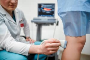
- Arterial Doppler ultrasound. This type of ultrasound assesses the circulation in a vein or artery. Doppler ultrasounds are often performed on the legs but can also be done on the arms or neck. This imaging can identify types of arterial blockage or injury to blood vessels, including blood clots and varicose veins. A vein or vascular specialist may use this type of ultrasound to measure the severity of varicose veins or to guide a venous procedure.
- Carotid ultrasound. A carotid ultrasound may also be called carotid duplex. The carotid arteries are located on each side of the neck. Using a small handpiece, the specialist can measure the amount of circulation happening through these arteries. Circulation here may be impaired due to the formation of arterial plaque, a sticky substance comprised of cellular debris, calcium, and fat. A buildup of plaque is what causes hardening of the arteries known as atherosclerosis.
- Renal artery ultrasound. The kidneys are the body’s filtration system that relies on robust circulation for optimal function. The renal arteries that supply nourishment to the kidneys can also become blocked or narrowed. Vascular ultrasound performed here may either diagnose or monitor a variety of conditions.
The Vascular Institute of Virginia has three convenient locations. Contact us at 703-763-5224 to schedule a visit in Woodbridge, Fredericksburg, or Fairfax, VA.