Inferior Vena Cava (IVC) Filter Placement
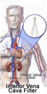
In an inferior vena cava filter placement procedure, interventional radiologists use image guidance to place a filter in the inferior vena cava (IVC), the large vein in the abdomen that returns blood from the lower body to the heart. The most common approach is to gain access to the jugular vein in the neck, using ultrasound guidance initially.
Blood clots that develop in the veins of the leg or pelvis, a condition called deep vein thrombosis (DVT), occasionally break up and large pieces of the clot can travel to the lungs. An IVC filter traps large clot fragments and prevents them from traveling through the vena cava vein to the heart and lungs, where they could cause severe complications or even death.
Inferior Vena Cava (IVC) Filter Placement/RemovalRemoval of an IVC Filter can be as easy as placing one. Using ultrasound guidance, the physician will access the jugular vein. In order to grab the filter, a snare or retrieval device is used to pull it out through the access sheath.
